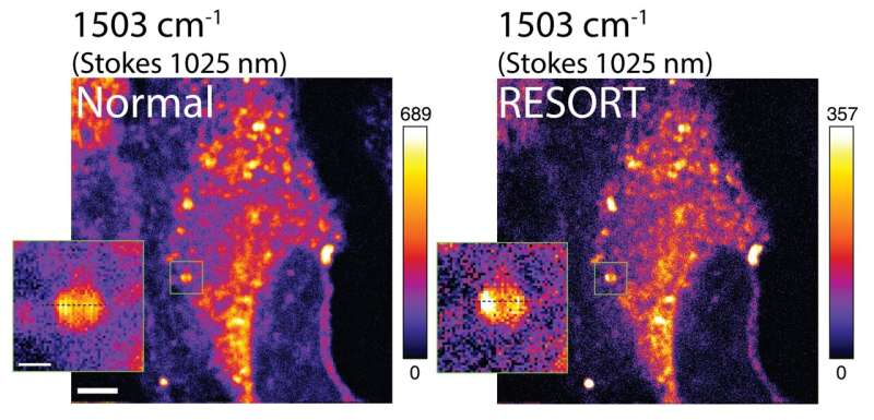
Advancements in imaging techniques have revolutionized the examination of biological samples at the microscopic level. Researchers from the University of Tokyo have taken a step further by combining elements from two leading imaging methods to create the RESORT imaging technique. This groundbreaking concept enables the observation of living systems with unparalleled detail. The results of this study can be found in the journal Science Advances.
Throughout history, optical devices have allowed us to explore the microscopic world with increasing precision. The more we can see, the more we can understand. Thus, there is a constant drive to improve our tools for examining the world around us, both externally and internally.
Modern microscopic imaging techniques have surpassed the capabilities of traditional microscopes. Two major technologies in this field are super-resolution fluorescence imaging, which offers high spatial resolution, and vibrational imaging, which allows for versatile labeling of various cellular components but compromises spatial resolution.
Professor Yasuyuki Ozeki from the University of Tokyo’s Research Center for Advanced Science and Technology explained the motivation behind the development of RESORT: “We aimed to overcome the limitations of existing imaging techniques and create something superior. RESORT, which stands for reversible saturable optical Raman transitions, combines the advantages of super-resolution fluorescence and vibrational imaging without the drawbacks of either. This laser-based technique utilizes Raman scattering, a unique interaction between light and molecules, to identify and analyze samples under a microscope. We successfully demonstrated RESORT imaging of mitochondria in cells, validating the effectiveness of this technique.”

The RESORT imaging process involves several stages, which, despite sounding complex, are less intricate than the techniques currently in use. First, the specific components of the sample to be imaged are labeled with photoswitchable Raman probes, which can control Raman scattering through the use of different laser lights employed by RESORT.
Next, the sample is placed within an optical setup that illuminates and captures an image of the sample. To facilitate this, the sample is irradiated with two-color infrared laser pulses for Raman scattering detection, ultraviolet light, and a donut-shaped beam of visible light. Together, these techniques confine Raman scattering to a specific area, allowing for highly precise imaging with exceptional spatial resolution.
According to Ozeki: “The goal is not just to obtain higher-resolution images of microscopic samples, as electron microscopes already excel in this area. However, electron microscopes tend to damage or disrupt the samples they observe. With RESORT, we are aiming to expand the palette of Raman probes to include more colors. This will allow us to capture the dynamics of various components within living samples, providing unprecedented insights into complex biological processes, disease mechanisms, and potential therapeutic interventions.”
While the primary focus of the research team was to enhance microscopic imaging in medical research and related fields, the laser design advancements made in the development of RESORT could have applications in other laser-related sectors that require high power or precise control, such as materials science.
More information:
Jingwen Shou et al, Super-resolution vibrational imaging based on photoswitchable Raman probe, Science Advances (2023). DOI: 10.1126/sciadv.ade9118. www.science.org/doi/10.1126/sciadv.ade9118
Citation:
Researchers create new imaging technique based on photoswitchable Raman probe (2023, June 16)
retrieved 16 June 2023
from https://phys.org/news/2023-06-imaging-technique-based-photoswitchable-raman.html
This document is subject to copyright. Apart from any fair dealing for the purpose of private study or research, no
part may be reproduced without the written permission. The content is provided for information purposes only.
Denial of responsibility! SamacharCentrl is an automatic aggregator of Global media. In each content, the hyperlink to the primary source is specified. All trademarks belong to their rightful owners, and all materials to their authors. For any complaint, please reach us at – [email protected]. We will take necessary action within 24 hours.

Shambhu Kumar is a science communicator, making complex scientific topics accessible to all. His articles explore breakthroughs in various scientific disciplines, from space exploration to cutting-edge research.

